At TheHealthBoard, we're committed to delivering accurate, trustworthy information. Our expert-authored content is rigorously fact-checked and sourced from credible authorities. Discover how we uphold the highest standards in providing you with reliable knowledge.
In Cardiology, what are P Waves?
In cardiology, P waves are basically graphic representations of the heart muscle’s atrial depolarization. They are part of a complex series of electrical waves that are detected during a non-invasive test of heart function called an electrocardiogram (ECG or EKG). Medical experts typically name the waves that show up on these tests according to alphabetical letters in order to quickly and succinctly identify them. Following this trend, the waves that directly follow P in an ECG are commonly known as the Q, R, S, and T waves. The P is the first deflection in the tracing of a heartbeat, typically seen as a small upward wave, and it is usually understood to be a representation of atrial depolarization. The first part usually looks like the rise of a small hill, and it indicates right atrial depolarization, while the far side, on the descent, represents left atrial depolarization. Doctors and other medical professionals use the depictions of these waves in diagnosing and treating certain heart conditions, and also as a way of monitoring a person or animal’s overall cardiovascular health.
Basics of Wave Representation

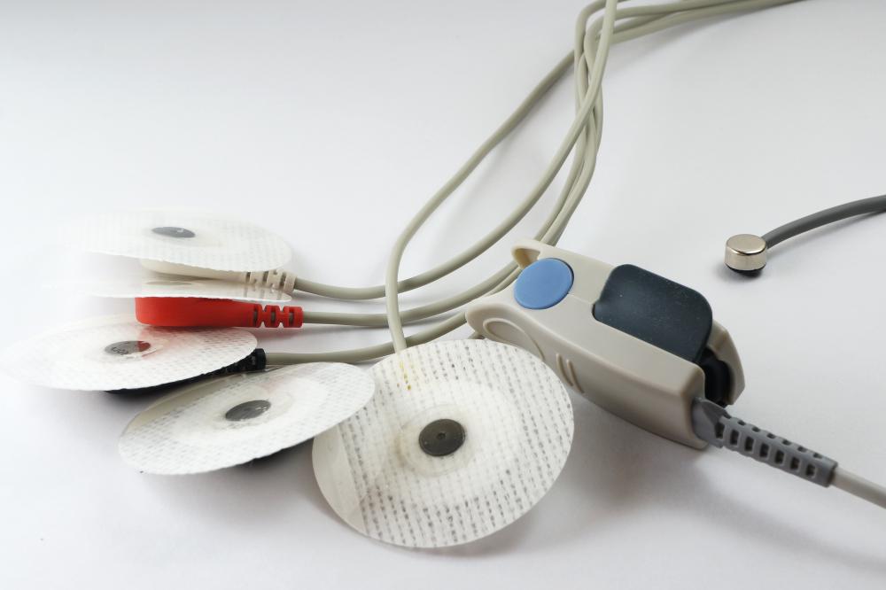
Electrical “polarization” and “depolarization” happen as a normal part of a heart’s beating. The beats pump blood through the internal cardiac chambers and into the arteries and veins of the whole body, but the heart is a lot more than just a pump: it’s also an electrical regulator, and its movements are kept in check by various charges and pulses that change slightly depending on exertion, blood chemistry, and strain. An ECG detects these subtle electrical changes and records them as a pattern of positive or negative waves on a digital monitor or on a paper readout. A physician or other healthcare professional can look at an ECG and see how the heart is functioning based on changes in the waves represented, and those with the “P” designation are part of the spectrum.

An ECG is sensitive enough to detect subtle electrical changes in the skin through monitoring leads placed near the heart. Depolarization of the heart muscle after a heartbeat, as it moves towards zero charge, activates the mechanisms in the heart that cause it to beat. A healthy heart will have a steady pattern of wave depolarization that can be seen on an ECG. A properly interpreted ECG can show the overall functioning of the heart. Specifically, the wave labeled P can indicate weakness or damage to the heart, both on its own and in relation to others.
What the Waves Should Look Like

Generally speaking, P waves should be no taller than 3 mm on a standard ECG. Taller waves may indicate right atrial enlargement, while longer waves that appear similar to the letter “m” may indicate left atrial enlargement. If an ECG shows multiple waves before the QRST complex, it may indicate second- or third-degree heart blockage.
P waves are important for many reasons, but when it comes to diagnostics their main benefit often has to do with timing: they almost always precede an atrial contraction, which typically occurs a fraction of a second later. On an ECG, P-style waves should appear as small upward-reaching hills. They should typically be of uniform height and a short distance from the QRST complex of waves. Changes in the shape, height, typical upward direction, or distance between the wave and the QRST complex can indicate heart problems.
How They Are Interpreted

It’s usually most common for all heart waves to be interpreted and studied by qualified cardiac health experts. These may not be the people actually performing the test at the outset, though. Depending on the circumstances, ECG technologists and medical aides may be the ones actually running the screenings and recording heart wave patterns. They don’t normally interpret these patterns, though. Once the waves have been charted, whether digitally or on paper print-outs, the results are usually sent to a heart expert who will study them and then discuss the findings with the patient.
Next Steps

Problems with waves in the P spectrum usually indicate a need for further care. Most of the time, doctors will order further testing before beginning treatment, though, in order to get a more certain read on what the problem really is. Abnormal waves in the P spectrum often indicate problems, but not always. They do give experts a narrower class of problems to screen for, however, which can make treatment and recovery more effective.
AS FEATURED ON:
AS FEATURED ON:


Discussion Comments
@NathanG - I agree that catheters are more precise; however it’s not true that the P waves are inconclusive. The article plainly states that they could indicate a right or left enlargement.
At least it will give you some idea of where to look. At any rate, these interpretations are best left up to the doctor not to the patient.
I am rarely given the readout of what my heart exam looks like, except if there is something wrong, in which case the doctor will explain what the results mean.
Now I know how to read the ECG report from the doctor. I have seen these printouts before but all it looked like was a bunch of waves with no rhyme or reason.
I am sure there is more to the report than just what the peaked P waves indicate, but at least this can give me an idea if there is an underlying problem. However, I think that even if a P wave indicates a problem with the heart, it’s probably not as decisive as say inserting a catheter or something like that.
To me, ECG diagrams are like static noise on the radio; they may give you a faint hint of a radio signal, but they don’t let you dial in exactly on what the problem is. That’s why the real test is with using catheters to inspect the blood vessels and determine if there is a blockage.
Post your comments