At TheHealthBoard, we're committed to delivering accurate, trustworthy information. Our expert-authored content is rigorously fact-checked and sourced from credible authorities. Discover how we uphold the highest standards in providing you with reliable knowledge.
In Cardiology, what are T Waves?
When the heart muscle alternately contracts and relaxes to pump blood, the electrical nerve waves that are producing those contractions spread throughout the body. This produces a characteristic pattern that can be measured by an electrocardiogram (ECG) when recorded from leads placed on the trunk or extremities. The T wave depicts the electrical wave accompanying relaxation of the ventricles, the two large pumping chambers on the bottom of the heart.
Doctors can look for alterations in the T wave, which is the most volatile of the various waves measured by an ECG, to spot potential problems with a patient's heart or general health. Abnormalities in the T wave may simply reflect a patient's advanced age, but many other conditions produce T wave disturbances. Damage in the area of the heart valves, reduced blood flow in the heart, and heart attacks may all cause T wave fluctuations.

Inflammation in or surrounding the heart, such as in myocarditis or pericarditis, may also contribute to T wave distortions. Physicians also note T wave changes following chest contusions with bruising or bleeding around the heart. Finally, some central nervous system problems, particularly hemorrhage into the lining membranes of the brain, cause a longer interval between the QRS waves and the T waves, which is called a long QT interval.

When physicians see flattened or inverted T waves, they may suspect poor blood flow to the heart. This also may occur when the left ventricle has increased in size due to sustained high blood pressure or atherosclerosis. Digoxin, a common cardiac drug, may produce this effect, as well. Abnormally tall T waves can be the first sign of a myocardial infarction or heart attack.

The QT interval exemplifies the time for both electrical waves that cause heart contraction and release to occur. On average, the QT interval lasts from 0.2 to 0.4 seconds. A prolonged QT interval can reveal some abnormally fast heart rhythms, called tachyarrthmias, which can lead to sudden death, while a short QT interval may indicate high blood calcium levels. The ST segment, which is the distance on the ECG from the S wave following the QRS wave to the peak of the T wave, indicates the length of time between maximal contraction of the ventricles and the beginning of its relaxation. In cases of poor ventricular blood flow, the ST segments may become elevated or depressed.
AS FEATURED ON:
AS FEATURED ON:

-
![T waves are displayed on an EKG graph.]() By: nyulT waves are displayed on an EKG graph.
By: nyulT waves are displayed on an EKG graph. -
![The T wave depicts the electrical wave accompanying relaxation of the ventricles, which are the two large pumping chambers on the bottom of the heart.]() By: stockshoppeThe T wave depicts the electrical wave accompanying relaxation of the ventricles, which are the two large pumping chambers on the bottom of the heart.
By: stockshoppeThe T wave depicts the electrical wave accompanying relaxation of the ventricles, which are the two large pumping chambers on the bottom of the heart. -
![The T wave is the most volatile of the various waves measured by an ECG.]() By: RTimagesThe T wave is the most volatile of the various waves measured by an ECG.
By: RTimagesThe T wave is the most volatile of the various waves measured by an ECG. -
![Inflammation in or surrounding the heart may contribute to T wave distortions.]() By: hrianaInflammation in or surrounding the heart may contribute to T wave distortions.
By: hrianaInflammation in or surrounding the heart may contribute to T wave distortions. -
![Abnormally tall T waves can be one of the first signs of a heart attack.]() By: Lisa F. YoungAbnormally tall T waves can be one of the first signs of a heart attack.
By: Lisa F. YoungAbnormally tall T waves can be one of the first signs of a heart attack. -
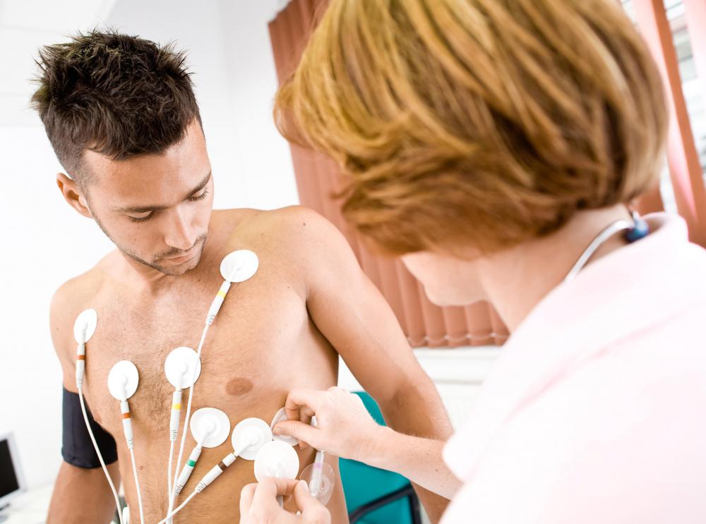
![During an electrocardiogram, electrodes are placed on the skin to monitor electrical impulses inside the heart.]() By: zayacskDuring an electrocardiogram, electrodes are placed on the skin to monitor electrical impulses inside the heart.
By: zayacskDuring an electrocardiogram, electrodes are placed on the skin to monitor electrical impulses inside the heart. -
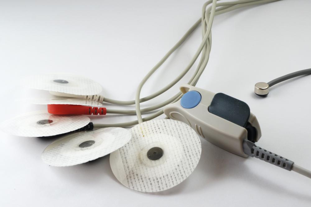
![An EKG is often used to continuously monitor the heart wave groups of a critically ill patient.]() By: sudok1An EKG is often used to continuously monitor the heart wave groups of a critically ill patient.
By: sudok1An EKG is often used to continuously monitor the heart wave groups of a critically ill patient.


Discussion Comments
This is great info. Thank you. I've been trying to get the doctors to explain this to me for weeks but felt like it was gibberish! It's so nice to actually be able to understand how this works. Thank you again. Another site added to 'bookmarks'.
Post your comments