At TheHealthBoard, we're committed to delivering accurate, trustworthy information. Our expert-authored content is rigorously fact-checked and sourced from credible authorities. Discover how we uphold the highest standards in providing you with reliable knowledge.
What are the Different Layers of Eye Tissue?
The human eye has three layers of eye tissue: the fibrous layer, the vascular layer, and the retina. Each of these layers performs a different function in helping a human see or in eye, as well as forms a place for muscles to attach to. The vascular layer protects portions of the eye. The fibrous layer of eye tissue allows light to enter the ourishes the eye and controls how much light comes into it. The retina is the section of the eye that allows a person to see.
The fibrous layer has two distinct parts. When a person sees the white of an eye, he is actually looking the sclera, a part of the fibrous layer of eye tissue. The sclera is white, opaque, and hard. It not only forms the shape of the eye but also provides a place for eye muscles to anchor. This layer helps to protect the rest of the eye.

At the front of the eye, the fibrous layer consists of the cornea, the place where light enters the eye. The cornea is the most exposed part of the eye and is filled with pain receptors. It is transparent and contains no blood vessels but is able to regenerate itself when it gets damaged. Since it has no blood vessels, it can be transplanted from person to person with little or no risk of rejection.

Much of the eye's pigment is located in the vascular layer of eye tissue. The pigment serves to absorb scattered light in order to prevent vision confusion. The choroid section of the vascular layer contains blood vessels that nourish the eye. The next layer, the ciliary body, forms a thick tissue around the lens. The ciliary body mostly contains muscles that control the lens's shape.

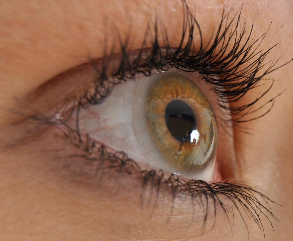
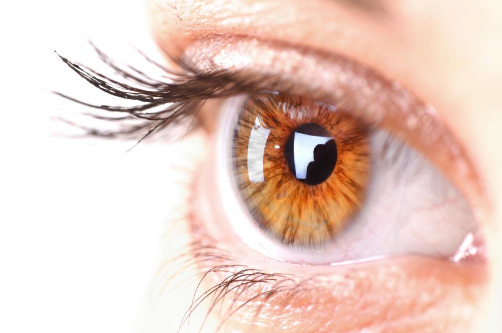
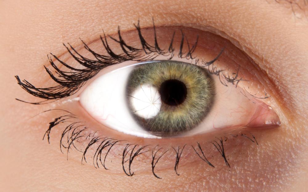
The iris is the part of the vascular layer of eye tissue that determines a person's eye color. Except for albinos, each person, regardless of the eye color, only possesses brown pigment in his eyes. People with brown eyes have pigment both at the back of the iris and in the body of the iris, and those with hazel eyes have less pigment in the body of the iris. If a person only has pigment at the back of the iris, the light can refract as it passes through the colorless part, allowing another person to only see a certain light wave. The result is the variety of different eye colors people may have, including shades of blue and green.

The pupil controls the amount of light that enters the eye. Muscles in the iris contract and dilate the pupil according to stress and the amount of light in the surrounding area. In a darkened room, when a person is scared, or when focusing on an object far away, the pupil will dilate to let in more light. If a person is in a bright room or is focusing on a nearby object, the pupil will contract to reduce the amount of light that enters the eye. Stress can also cause the pupil to dilate.

The eye tissue known as the retina — the part of the eye that allows a person to see — is separated into two layers. The outer layer, the pigmented layer, absorbs light, as well as removes damaged and dead photoreceptor cells. It also serves to recycle the vitamin A product that eyes need to see. The inner layer, the nueral layer, contains the photoreceptors and other cells that allow a person to see. When light hits them, they generate a signal that is sent to the brain and is translated as vision.
AS FEATURED ON:
AS FEATURED ON:

-
![Hazel eyes contain less pigment in the body of the iris than brown eyes.]() By: Dan RaceHazel eyes contain less pigment in the body of the iris than brown eyes.
By: Dan RaceHazel eyes contain less pigment in the body of the iris than brown eyes. -
![The cornea is the transparent portion of the eye which allows light to enter.]() By: kocakayaaliThe cornea is the transparent portion of the eye which allows light to enter.
By: kocakayaaliThe cornea is the transparent portion of the eye which allows light to enter. -
![People with brown eyes have pigment at the back of the iris and in the body of the iris.]() By: katalinksPeople with brown eyes have pigment at the back of the iris and in the body of the iris.
By: katalinksPeople with brown eyes have pigment at the back of the iris and in the body of the iris. -
![The sclera, or white part of the eye, is part of the fibrous layer of eye tissue.]() By: Péter MácsThe sclera, or white part of the eye, is part of the fibrous layer of eye tissue.
By: Péter MácsThe sclera, or white part of the eye, is part of the fibrous layer of eye tissue. -
![Regardless of eye color, people possess brown pigment in their eyes.]() By: arturas kerdokasRegardless of eye color, people possess brown pigment in their eyes.
By: arturas kerdokasRegardless of eye color, people possess brown pigment in their eyes. -
![The six extraocular muscles are the lateral, medial, inferior, and superior rectus, and the inferior and superior oblique muscles.]() By: blueringmediaThe six extraocular muscles are the lateral, medial, inferior, and superior rectus, and the inferior and superior oblique muscles.
By: blueringmediaThe six extraocular muscles are the lateral, medial, inferior, and superior rectus, and the inferior and superior oblique muscles.


Discussion Comments
I have chronic dry eye syndrome, and I was not aware of the different layers of eye tissue until my eye doctor explained them to me. I had no idea that keeping the cornea moist helps protect it from injury and infection. I have to use artificial tears and ointment at night to keep the cornea properly lubricated.
Post your comments