At TheHealthBoard, we're committed to delivering accurate, trustworthy information. Our expert-authored content is rigorously fact-checked and sourced from credible authorities. Discover how we uphold the highest standards in providing you with reliable knowledge.
What is a Brain Scan?
A brain scan is an image of the brain that can be obtained by specific types of X-rays. When a medical professional orders a scan, he may be looking for abnormalities, such as a stroke or a brain tumor. He determines who needs this type of X-ray based on the the symptoms the individual is experiencing. There are two main types: computed tomography (CT) and magnetic resonance imaging (MRI).
The CT scan is usually the first type of image that is ordered if an individual presents to the emergency room with symptoms of a stroke. During a CT, the individual will lie on a table, which will slide into a large circular tube. The interior of the scanner rotates around the individual to take pictures of the entire brain. Sometimes, the healthcare professional may order a contrast material injected into the vein to help highlight abnormal areas in the brain.

These images can be viewed by the medical professional in "slices" to detect any abnormalities. The brain on a CT scan will look gray, and any abnormalities will show up as darker or white areas in the brain or surrounding tissue. The type of stroke that an individual experiences can usually be determined by the CT brain scan.
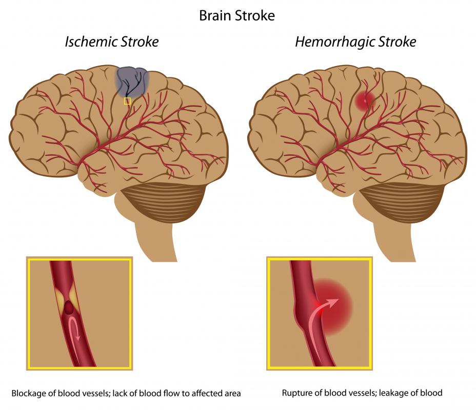
There are two types of strokes. One type is called an ischemic stroke, which occurs when blood supply to the brain is blocked, and causes a part of the brain to die. An ischemic stroke area will appear darker than the normal brain color on the CT scan. A hemorrhagic stroke occurs when a blood vessel in the brain ruptures and bleeding into the brain occurs. The blood in the brain will show up as a white area on the CT scan. There are times, however, when the CT scan cannot detect a stroke right away, and sometimes the area of the brain affected by the stroke will not appear abnormal for several hours.

The CT scan is the first type of X-ray performed in the emergency room. If a definite diagnosis cannot be made from this test, then an MRI scan is usually ordered. The MRI can take a more detailed picture of the brain. It uses magnets, along with radio waves, to transport an image of the inside of the brain to a computer screen.
When an MRI is being performed, the individual will lie on a table and the table will be slid into a long tunnel. The machine will take pictures of the brain from all angles. The MRI produces a more detailed image, so these pictures can provide a much clearer image of the soft tissue of the brain. It can often detect abnormalities that the CT scan cannot.
AS FEATURED ON:
AS FEATURED ON:

-
![A full MRI scan often includes several different layouts of the brain, offering a complete look at the patient's neurological health.]() By: Marcin SadlowskiA full MRI scan often includes several different layouts of the brain, offering a complete look at the patient's neurological health.
By: Marcin SadlowskiA full MRI scan often includes several different layouts of the brain, offering a complete look at the patient's neurological health. -
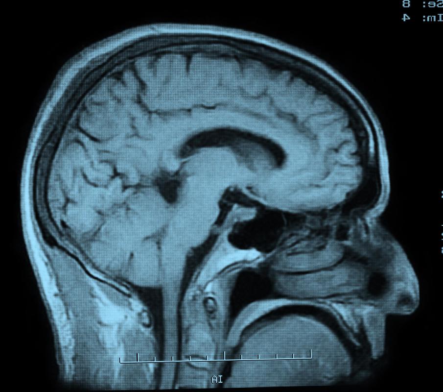
![An MRI produces a very detailed image of the brain.]() By: Monkey BusinessAn MRI produces a very detailed image of the brain.
By: Monkey BusinessAn MRI produces a very detailed image of the brain. -
![CT scans are automatically ordered if an individual has shown signs of a stroke, or similar neurological ailments.]() By: Katrina BrownCT scans are automatically ordered if an individual has shown signs of a stroke, or similar neurological ailments.
By: Katrina BrownCT scans are automatically ordered if an individual has shown signs of a stroke, or similar neurological ailments. -
![The human brain is subject to a number of severe ailments, the most common of which is a stroke.]()
The human brain is subject to a number of severe ailments, the most common of which is a stroke. -
![A diagram of an ischemic stroke and a hemorrhagic stroke.]() By: AlilaA diagram of an ischemic stroke and a hemorrhagic stroke.
By: AlilaA diagram of an ischemic stroke and a hemorrhagic stroke. -
![An image of the brain can be obtained by specific types of X-rays.]() By: CGinspirationAn image of the brain can be obtained by specific types of X-rays.
By: CGinspirationAn image of the brain can be obtained by specific types of X-rays. -
![A functional MRI allows the radiologist to see brain processes, and must be interpreted in real time.]() By: EPSTOCKA functional MRI allows the radiologist to see brain processes, and must be interpreted in real time.
By: EPSTOCKA functional MRI allows the radiologist to see brain processes, and must be interpreted in real time. -
![When a medical professional orders a scan, he may be looking for a brain tumor.]() By: rob3000When a medical professional orders a scan, he may be looking for a brain tumor.
By: rob3000When a medical professional orders a scan, he may be looking for a brain tumor. -
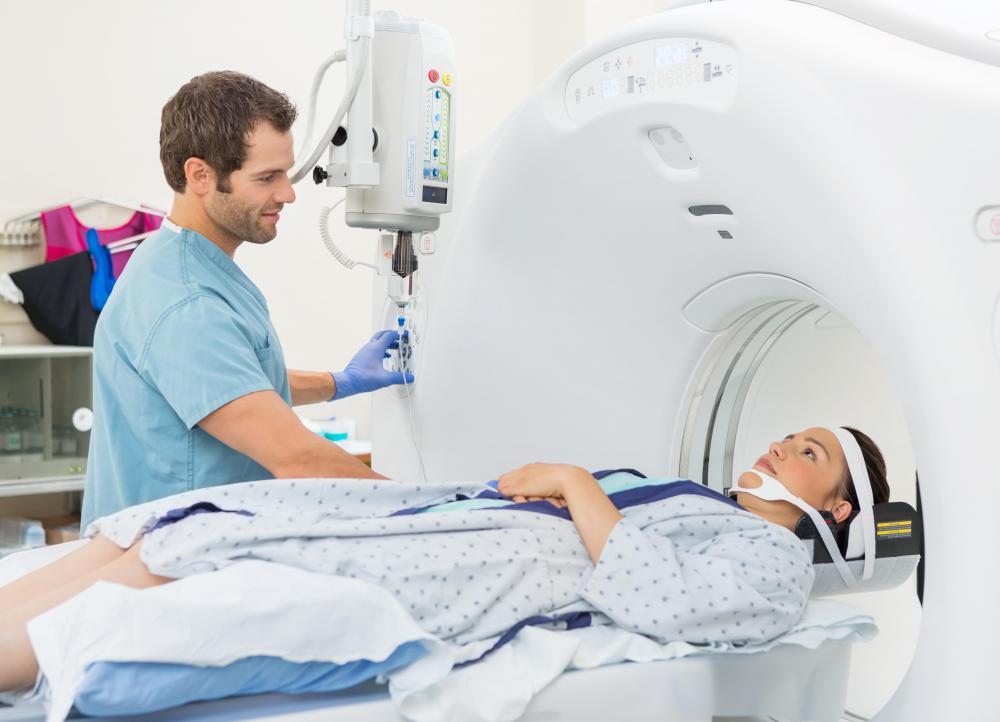
![CT scans and MRIs are used to take images of the brain from multiple angles.]() By: Tyler OlsonCT scans and MRIs are used to take images of the brain from multiple angles.
By: Tyler OlsonCT scans and MRIs are used to take images of the brain from multiple angles.


Discussion Comments
If the brain scan shows a stroke, what's the treatment?
Post your comments