At TheHealthBoard, we're committed to delivering accurate, trustworthy information. Our expert-authored content is rigorously fact-checked and sourced from credible authorities. Discover how we uphold the highest standards in providing you with reliable knowledge.
What Is a Sacrococcygeal Teratoma?
A sacrococcygeal teratoma (SCT) is a tumor that is most commonly seen in infants and young children. This tumor represents a proliferation of a number of different types of cells. It is commonly identified after finding a lump or bump in the region of the lower back, and is diagnosed with different imaging tests. All SCTs should be surgically removed, and some affected patients will also require chemotherapy in order to adequately treat this condition.
A teratoma is an abnormal growth of germ cells. The early development of embryos depends on these germ cells, which are undifferentiated cells that grow and develop into all of the components of the human body. Since teratomas develop from these germ cells, they can contain a variety of different types of tissue including glands, bones, teeth, hair, or skin. The term sacrococcygeal refers to a region of the body; the sacral region is the lower back, and the coccygeal region is the tailbone area. Therefore, a sacrococcygeal teratoma is a tumor comprised of many different types of cells that is located in the region of the lower back and tailbone.

Sacrococcygeal teratomas can cause a variety of symptoms, the most common being a mass protruding from the sacrococcygeal region of the body. Other symptoms can develop from the internal growth of the tumor, which can cause blockage of the urinary tract or the gastrointestinal tract. Obstruction of the urinary system can cause decreased urination, blood in the urine, or kidney failure. Blockage of the gastrointestinal tract can result in bowel obstruction, which causes symptoms such as abdominal distension, vomiting, constipation, and pain.

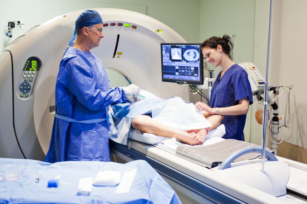
The diagnosis of sacrococcygeal teratoma can be made by a number of methods. Often, SCTs can be diagnosed by prenatal ultrasound. The diagnosis is confirmed in these patients after birth by imaging studies such as computed tomography (CT) scans or magnetic resonance imaging (MRI) scans. Infants and young children can also be diagnosed with SCTs. These patients are identified based on their symptoms, and the diagnosis of SCT is confirmed with a CT or MRI.

The first step in the treatment of a sacrococcygeal teratoma is surgery, which is necessary because the tumor could be malignant. The method used to perform the surgery depends on the size of the SCT. Often the tumors can be very large, and the scope of surgery can be extensive. Sometimes multiple surgeries might be required, and in some cases removal of the coccyx might be necessary.

After being surgically removed, the sacrococcygeal teratoma is sent to a pathologist for evaluation. The pathologist will determine whether the tumor is benign or malignant. If the tumor is benign, often no additional treatment is required other than following up with the patient to monitor for recurrence. Unlike benign tumors, patients found to have malignant tumors will require chemotherapy in order to fully eradicate any abnormal cells that might still be present in the body.
AS FEATURED ON:
AS FEATURED ON:

-
![Many cases of SCT can be diagnosed via ultrasound.]() By: Mikael DamkierMany cases of SCT can be diagnosed via ultrasound.
By: Mikael DamkierMany cases of SCT can be diagnosed via ultrasound. -
![A pathologist can determine whether a sacrococcygeal teratoma is malignant or benign.]() By: nandyphotosA pathologist can determine whether a sacrococcygeal teratoma is malignant or benign.
By: nandyphotosA pathologist can determine whether a sacrococcygeal teratoma is malignant or benign. -
![Diagnosis of sacrococcygeal teratoma may be achieved through MRI scans.]() By: EPSTOCKDiagnosis of sacrococcygeal teratoma may be achieved through MRI scans.
By: EPSTOCKDiagnosis of sacrococcygeal teratoma may be achieved through MRI scans. -
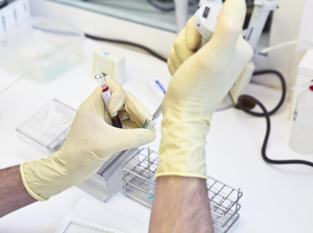
![A pathologist may use various methods to determine the chemical composition of germ cells.]() By: spflaumA pathologist may use various methods to determine the chemical composition of germ cells.
By: spflaumA pathologist may use various methods to determine the chemical composition of germ cells. -
![Patients undergoing chemotherapy for sacrococcygeal teratoma may have a port placed under the skin in order to accommodate the need for frequent venous access.]()
Patients undergoing chemotherapy for sacrococcygeal teratoma may have a port placed under the skin in order to accommodate the need for frequent venous access. -
![Obstruction of the urinary system as a result of sacrococcygeal teratomas may cause kidney failure.]() By: Sebastian KaulitzkiObstruction of the urinary system as a result of sacrococcygeal teratomas may cause kidney failure.
By: Sebastian KaulitzkiObstruction of the urinary system as a result of sacrococcygeal teratomas may cause kidney failure. -
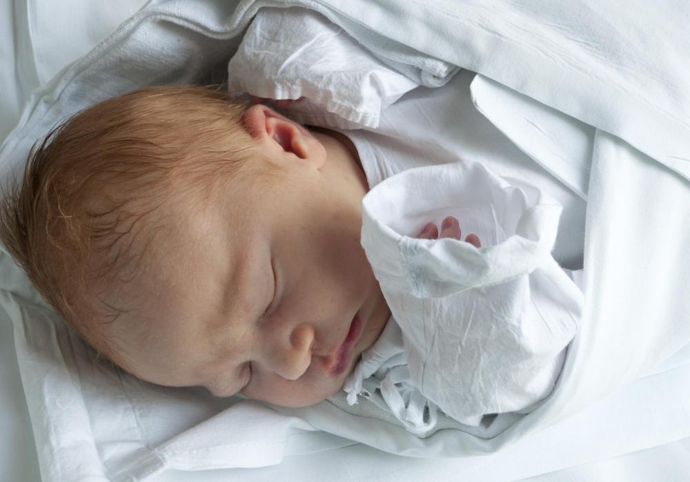
![Sacrococcygeal teratomas are most commonly seen in infants and small children.]() By: jamdesignSacrococcygeal teratomas are most commonly seen in infants and small children.
By: jamdesignSacrococcygeal teratomas are most commonly seen in infants and small children.


Discuss this Article
Post your comments