At TheHealthBoard, we're committed to delivering accurate, trustworthy information. Our expert-authored content is rigorously fact-checked and sourced from credible authorities. Discover how we uphold the highest standards in providing you with reliable knowledge.
What is a Sonogram?
A sonogram is a medical procedure that uses ultrasound waves to create a picture of something that is happening within a person’s body. This is a very common procedure in pregnancy, and is what produces the black-and-white fetal pictures that so many new parents proudly display to friends and family. Medical professionals use the technology for a range of conditions, however, including cancer biopsies and organ evaluations. The pictures generated from sonograms give experts a relatively clean look into the body to understand what is happening without having to perform surgery or other invasive procedures.
How it Works

Sonogram machines emit sound waves, often known as ultrasound waves, that bounce off of organs, bones, and muscles. The machines are able to calculate the distance between waves in order to generate a very accurate picture, which is displayed on a specialized computer screen.
In most cases, the waves are both sent and received from a wand-like instrument known as a transducer. A trained technician will usually gently touch the wand to the skin above the area of interest. Jelly or lubricant is often applied first, both to help the wand glide and to amplify the sound waves to generate a clearer, more accurate picture. Most sonogram sessions result in pictures from many different angles, which technicians and medical professionals look at in sequence to get an idea of what is happening internally.
Uses in Pregnancy

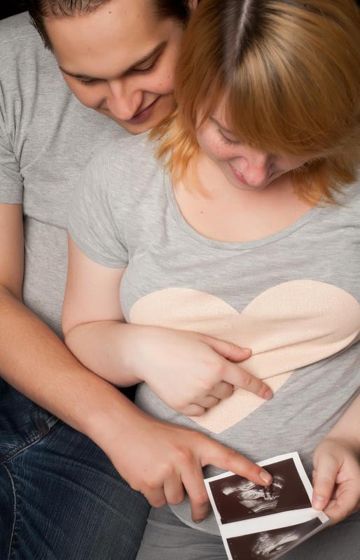
Doctors and medical teams in many parts of the world use sonogram technology as a way to monitor pregnancy. Sonogram pictures can help identify the basic anatomy of a developing fetus, and can often detect birth defects and abnormalities very early on. Most of the time, this technology can also identify the gender of the growing child.
Standard sonogram transducers cannot always detect the signs of a very early pregnancy. When there is reason to check on fetal progress within the first few months of development, medical experts must usually use what is known as a transvaginal transducer, a very narrow wand that must be inserted into the pregnant woman’s vagina. Once in place, the transducer is usually able to detect the presence of fetal growth — and can often even capture the baby’s heartbeat.
Other Medical Uses

Healthcare practitioners commonly use ultrasound technology to investigate unknown causes of internal pain as well as to diagnose and monitor unusual growths such as tumors. The images produced from the sound waves can help experts get a clear visual of what is happening inside the body. The technology can identify organ malformations, bruising, or other internal injuries. Getting a good picture before surgery or other treatment can help medical professionals make the best, most appropriate recommendations.
Possible Risks

Sonograms are widely considered low-risk, and are typically classified as “non-invasive.” This does not mean that they are always entirely safe, however. The sound waves used have been known to heat body tissues slightly, and can also create small pockets of gas on rare occasions. While not necessarily harmful, these effects will occasionally cause complications, particularly when blood vessels and bone density are involved — heated gasses in these areas can cause discomfort, blood clotting, and structural weakening.

To mitigate the possible risks to a developing child, most medical professionals use sonograms only intermittently in pregnancy. Though parents often want to see images of their growing baby at each doctor visit, this is not usually permitted unless there is a legitimate medical need. Limiting the number of sonograms administered is one of the ways that medical professionals protect babies from potential risks. Listening to the fetal heartbeat and monitoring the mother’s blood work are often just as effective at making sure the baby stays healthy as a real-time picture is.
Preparation for the Procedure
Sonograms are typically very easy to perform, and patients do not usually need to do anything special to prepare. Wearing loose-fitting clothing is usually a good idea, as technicians will often need to shift or move garments around in order to place the transducer on the skin. Otherwise, just remaining calm and taking deep breaths is usually the best way to get ready.
AS FEATURED ON:
AS FEATURED ON:

-
![A sonogram shows the basic anatomy of a developing fetus.]() By: Oleksandr BondarA sonogram shows the basic anatomy of a developing fetus.
By: Oleksandr BondarA sonogram shows the basic anatomy of a developing fetus. -
![A diagnostic ultrasound uses high-frequency sound waves to create an image of the inside of a body.]() By: poco_bwA diagnostic ultrasound uses high-frequency sound waves to create an image of the inside of a body.
By: poco_bwA diagnostic ultrasound uses high-frequency sound waves to create an image of the inside of a body. -
![A sonogram photo is often a cherished keepsake for expectant parents.]() By: DNF-StyleA sonogram photo is often a cherished keepsake for expectant parents.
By: DNF-StyleA sonogram photo is often a cherished keepsake for expectant parents. -
![A sonogram may be used to identify the gender of an unborn baby.]() By: Michael IrelandA sonogram may be used to identify the gender of an unborn baby.
By: Michael IrelandA sonogram may be used to identify the gender of an unborn baby. -
![Sonograms can offer views of an unborn child.]() By: Monkey BusinessSonograms can offer views of an unborn child.
By: Monkey BusinessSonograms can offer views of an unborn child.


Discussion Comments
The safety of these tests are often contingent upon the technician. In my case, I wasn't so lucky as the tech dug the transducer deep into my hernia patch and plug and dislodged it, causing a painful hernia recurrence. I can barely walk and the pain ranges from a 3 to a 7. To get surgery and mesh removal would be brutal since it has been in me for 15 years and has incorporated into my anatomy and no surgeon wants to touch me!
I found Vonmajor's article to be very interesting, helpful and full of intelligent facts and common sense. My daughter is pregnant now (early stage - so less worry per this article) and the picture is very clear! More clear than MRI x-rays. Is it because it's closer to the skin in a sonogram and if so, can it be used to see inside knee injuries or elbows, especially torn ligaments, tendons, etc.? --Sherry
This is a completely misleading article to state that sonograms are completely safe. Ultrasound does too, like x-ray that predated the instrument, have ionizing radiation. Appreciably less, but nonetheless it has ionizing radiation. It also induces heat in the tissue to the embryo/fetus.
Early gestation embryos have heat shock proteins to protect themselves from heat to a degree, but later the fetus does not. Babies are seen to recoil away from the source of the device, and deaths have occurred in some rare cases immediately after the use of a ultrasound.
Where controls have been used, as in animal studies, ultrasound in gestation in cats, mice and monkeys has shown decided behavioral effects on the offspring. Upon examination of neural tissue of the lab animals above, brain damage in the same areas that is similar to that found in humans who have autism symptom disorders. These lab results have been observed when only two ultrasounds have been delivered during gestation.
One of the biggest mysteries today is the tremendous rise in the incidence of autism. Some forty years ago it was classified as 1 in 10,000 births. Now it is 1 in 88. Are vaccines the cause? Much of the thimersol (mercury as a preservative )in vaccines was removed to try to mitigate the rise. Nothing changed. The rate continued to rise. Meanwhile, the use and frequency of ultrasounds has taken off during the same period.
I work in radiation health protection. Federal regulations stipulate that a declared pregnant woman is to be limited to 10 percent of the ionizing radiation limit of an adult based upon fetal sensitivity. The most sensitive tissues in the body are the bone marrow, reproductive and the lining of the small intestine. These tissues in a baby in development are very sensitive. Could it be that a very common medical condition of those with autism and Down syndrome is a very high prevalence of digestive problems? Could bombardment of this sensitive tissue during the most critical developmental and delicate stage of be a significant source of the above mentioned conditions?
In our field, we use a mean of measurements such and millisieverts, or millirem as units of exposure. Ask your practitioner what degree of exposure they are giving to your baby. They probably do not have a unit. They will just try to placate you and tell you it is safe. Question them further and ask them to what threshold of exposure is safe and I doubt if they can give you a unit or value.
Furthermore, are all devices calibrated to deliver a uniform exposure, or do they just judge it upon picture quality? It is like giving a person a hammer. Pretty soon, all the world looks like a nail ready to be hammered upon. It’s the same with the sonogram. It can be useful, but only in limited instances. Plus, 80 to 90 percent of the risks involved in terms of birth defects can be determined by passive blood tests. Unless there are serious complications and you have to see inside, the procedure is not medically sound enough to justify the extra risks that appear to be manifesting themselves. We are highly encouraged to have a questioning attitude in my profession. For the health of our babies it applies even more. Do not accept the phrase, “Trust us.” --Jerry
yes you can hear the heart beat through a sonogram!
would a female baby in the mother's womb show the ovaries.
can a sonogram tell how many weeks pregnant you are?
We have a mentally challenged woman who doesn't speak. She is in severe pain in her stomach. The wait time for a sonogram is eight months. Is there any other way?
grandson diagnosed with mrsa in blood. will a sonogram tell if he has infection in his heart? can he be cured?
I heard if all is normal, not to have more than two sonograms. Been told its hard on baby's ears. Any true to this?
my doctor order a sonogram for me because of my severe bleeding due to an IUD. can the sonogram detect why am bleeding so severely.
would a sonogram find out why my mother is losing so much blood all the time? She has had gastroscopy and endoscopy and they found nothing.
My doctor ordered a sonogram because of a mass on my back. She also ordered a biopsy. Can this really be that I have cancer?
How soon till a pregnancy show up on a sonogram?
You can hear the heart beating during a sonogram if the machine has doppler controls.
"The Neighbor" has been using some sonographic tool to harass and maliciously abuse me and my pets, including plants; my arrowhead plant was desiccated in perfect preservation, perfect greenness, and looked alive, but was powder dry and crumbled to dust when I touched it. The dogs have cataracts, and water I leave out for them dehydrates very very quickly. I have to constantly give them water. As myself. I don't purchase glasses, because they become brittle and shatter. This has been going on for a while. What can I do about this? Local "law enforcement" seem to think it's a joke. I'll check back to your site to see if you respond.
All the references I've seen to sonogram indicate the wonderful health benefits, benefits I'm sure are real. But being abused 24/7 is insane. I've already seen what it did to my plant/s, the dogs, how about me? Brain damage? It causes heat exhaustion/stroke in myself and the dogs.
The common misconception about sonograms is that the most common use is to monitor a pregnancy. The most common use of sonograms in hospitals and clinics worldwide is for diagnostic medical purposes, hence, the letters after a registered sonographer's name, R.D.M.S. = Registered Diagnostic Medical Sonograper. A sonographer can be registered in Obstetrics and Gynecology, Abdomen, Breasts, Vascular, Echocardiography, etc. Ultrasound is also used to guide many biopsy procedures as well as paracentesis and thoracentesis and has many other diagnostic uses. The sonographer is often the "doctor's eyes" in diagnosing pathology.
how safe are those 3D and 4D sonograms, and do you think that they'll become the norm for sonograms, replacing the black and white versions?
Is a sonogram effective in examining the prostate for cancer? And if it is, is it as effective as a biopsy?
Can you listen to the heartbeat through a sonogram?
Post your comments