At TheHealthBoard, we're committed to delivering accurate, trustworthy information. Our expert-authored content is rigorously fact-checked and sourced from credible authorities. Discover how we uphold the highest standards in providing you with reliable knowledge.
What is Scleritis?
Scleritis is an inflammation of the white section of the outer layer of your eyeball, also known as the sclera. There are a variety of forms of the condition, with a range of possible severe symptoms. It's usually a treatable condition, but can be connected to underlying problems.
The sclera is a protective coating for your eye, made up of connective tissue. Most people know it simply as the white of the eye, though the color can vary slightly in children and elderly people. As well as maintaining the round shape of the eye, it protects much of the inner working parts of the eye.

In rare cases, scleritis affects the back of the eye, a condition known as posterior scleritis. However, most cases are of anterior scleritis, affecting the part of the sclera which is visible. The condition usually involves persistent reddening of the affected section, accompanied by a pain which can either be constant or pulsating.
The most common form is known as diffuse anterior scleritis. More advanced cases may involve nodular anterior scleritis in which individual tiny red swellings appear. This has around a 20 percent risk of developing into necrotising scleritis, the most severe form. Otherwise known as brawny scleritis, this involves severe inflammation to the point that the sclera become thinner.

In some cases, thinning of the sclera can lead to it forming into bulges. In severe cases the sclera thins so much that the eyeball is at risk of puncturing. Other risks from scleritis include damage to the cornea, which is the protective transparent material in front of the iris and pupil. Scleritis can also lead to damage to the back of the eye.
Generally scleritis develops as the result of underlying conditions. These are usually related to connective tissue or autoimmune diseases. The most common conditions behind scleritis are rheumatoid arthritis and Ankylosing spondylitis.
Scleritis is usually diagnosed by a physical examination by an eye specialist. CT scans and MRI scans can help detect some cases. However, most of the evidence of scleritis is easily visible so scans are not usually needed.

In many cases, scleritis is treatable simply by using non-steroidal anti-inflammatory drugs to reduce pain and inflammation. This will remove the symptoms, but the underlying problem which led to scleritis will still need to be dealt with. In severe cases, either steroids or immunosuppressive drugs may be required to treat the scleritis.
AS FEATURED ON:
AS FEATURED ON:

-
![An MRI scan may help detect scleritis.]() By: Mikhail KondrashovAn MRI scan may help detect scleritis.
By: Mikhail KondrashovAn MRI scan may help detect scleritis. -
![Scleritis is usually diagnosed through a physical examination by an eye specialist.]() By: Peter Orsaeo SrScleritis is usually diagnosed through a physical examination by an eye specialist.
By: Peter Orsaeo SrScleritis is usually diagnosed through a physical examination by an eye specialist. -
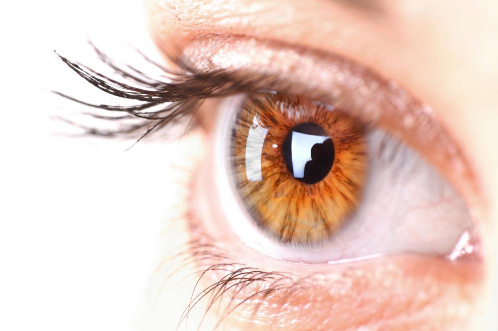
![Scleritis is the inflammation of the white part of the eye, commonly called the sclera.]() By: Péter MácsScleritis is the inflammation of the white part of the eye, commonly called the sclera.
By: Péter MácsScleritis is the inflammation of the white part of the eye, commonly called the sclera. -
![Risks from scleritis include damage to the cornea, iris, and pupil.]() By: stockshoppeRisks from scleritis include damage to the cornea, iris, and pupil.
By: stockshoppeRisks from scleritis include damage to the cornea, iris, and pupil.


Discuss this Article
Post your comments