At TheHealthBoard, we're committed to delivering accurate, trustworthy information. Our expert-authored content is rigorously fact-checked and sourced from credible authorities. Discover how we uphold the highest standards in providing you with reliable knowledge.
What is the Anatomy of the Leg?
The anatomy of the leg consists of those parts of the lower limb between the knee and the ankle. It features two bones known as the tibia, or shin bone, and the smaller fibula. Depending on their location in the anatomy of the leg, its muscle groups are divided into four different regions called compartments. The muscles of the leg bend the foot, assist in walking, and are necessary for coordinated movements like those in sports.
The tibia is the much thicker bone of the leg. This is because it transfers all of the weight from the femur above it to the ankle and foot. After the femur, it's the strongest and largest bone of the body. At the upper end, it has three points, called condyles, which meet with the femur. At the lower end, it meets the talus bone and flares out at the ankle on the inside of the leg. The body of the tibia appears triangular in shape.

The fibula, the rear or posterior bone in the anatomy of the leg, doesn't support any of the body's weight. Still, it serves as a point for the attachment for several muscles. It is connected to the tibia along their length by a band of tissue known as an interosseous membrane. It also joins the tibia near both the knee and ankle.
There are four main muscle groups in the anatomy of the leg. At its front, major muscles in the anterior compartment include the tibialis anterior and the extensor digitorum longus. All the muscles of the anterior compartment assist in flexing the foot back toward the leg. These muscles can become inflamed in the painful condition known as shin splints.

Muscles of the lateral compartment, on the inside of the leg, include the fibularis longus and the fibularis brevis. These muscles move the foot from side to side and also flex the foot downward. At the back of the leg, the posterior group is divided into superficial and deep posterior compartments. Together these groups help to flex the ankle downward. They also have a number of functions in various forms of movement, including walking, running, and various athletic movements.
The muscles of the leg receive blood mostly through the anterior and posterior tibial arteries. They supply the leg muscles with blood as they branch along the tibia and fibula. The leg is innervated by the sciatic nerve.
In contrast to the usual use of the term, the anatomy of the leg refers strictly to those parts between the ankle and knee. The lower limb is divided into the thigh, ankle, and foot. The thigh extends from the hip to the knee.
AS FEATURED ON:
AS FEATURED ON:

-
![A person with defined leg muscles.]() By: unpictA person with defined leg muscles.
By: unpictA person with defined leg muscles. -
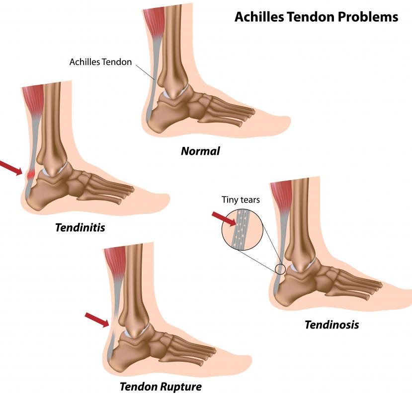
![This diagram shows some common problems with the Achilles tendon, part of the anatomy of the leg.]() By: AlilaThis diagram shows some common problems with the Achilles tendon, part of the anatomy of the leg.
By: AlilaThis diagram shows some common problems with the Achilles tendon, part of the anatomy of the leg. -
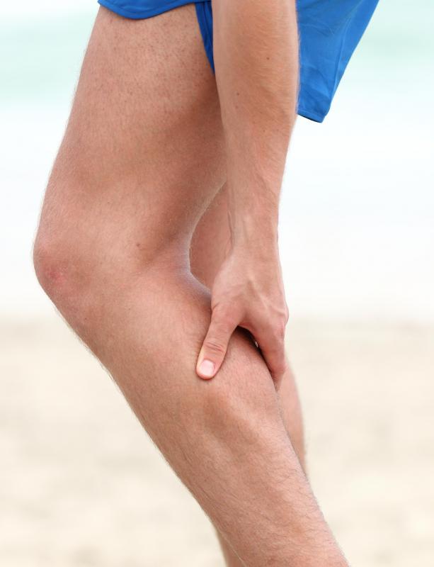
![A man with a calf cramp.]() By: MaridavA man with a calf cramp.
By: MaridavA man with a calf cramp. -
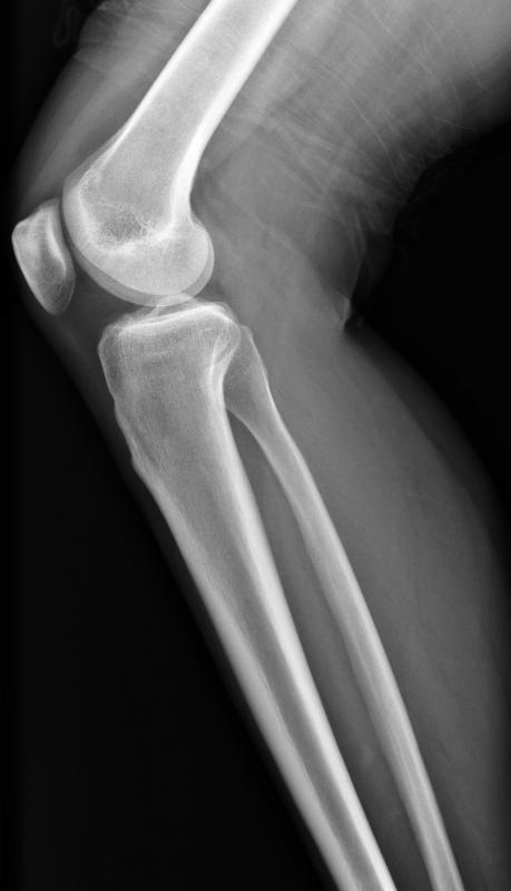
![This x-ray shows the femur, patella (kneecap), tibia, and fibula.]() By: itsmejustThis x-ray shows the femur, patella (kneecap), tibia, and fibula.
By: itsmejustThis x-ray shows the femur, patella (kneecap), tibia, and fibula. -
![The bone in the upper part of the leg is called the femur or thighbone.]() By: maya2008The bone in the upper part of the leg is called the femur or thighbone.
By: maya2008The bone in the upper part of the leg is called the femur or thighbone.


Discuss this Article
Post your comments