At TheHealthBoard, we're committed to delivering accurate, trustworthy information. Our expert-authored content is rigorously fact-checked and sourced from credible authorities. Discover how we uphold the highest standards in providing you with reliable knowledge.
What Is the Anatomy of the Skeletal Muscles?
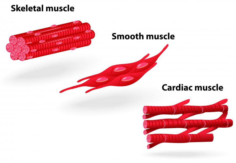
Despite the vast array of different muscles that attach and move the skeletal structure, the anatomy of the skeletal muscles is basically the same throughout the body. Skeletal muscles are made up of long multinucleate cells that are cylindrical in shape. The muscles are almost always attached directly to the bones, and unlike the other two muscles types, cardiac and smooth, are under voluntary control. The skeletal muscles have many layers and membranes that protect and compartmentalize their various components. The multinucleate cells may also be called fibers that group together in bundles depending on the voluntary action for which they control.
The first layer of the skeletal muscles is the thin, elastic-like membrane called the sarcolemma that covers the individual cells. The main function of the sarcolemma is to keep all the various cell components intact, in much the way a cell membrane functions in other cells of the body. Within the sarcolemma, the muscle cell’s fluid, or sarcoplasm, contains pl,the myofibril. Each individual fiber contains numerous myofibrils, which are protein strands that run the whole length of the muscle fiber. These structures lay side-by-side and are responsible for lengthening and contracting the muscle at the signal of the central nervous system (CNS).

Myofibrils can be further dissected into another component that is more directly involved in the contraction and lengthening of the fiber, the sarcomere. Sarcomeres act as tiny chains of contractile tissue that are aligned end-to-end throughout the myofibril. At the microscopic level, the myofibril is composed of an even finer strand of protein called a myofilament. Myofilament proteins are composed of dark strands, or anisotropic (A) bands, and light strands, or isotropic (I) bands; the contrasting colored proteins are what give skeletal muscle its striated appearance. The A-bands and I-bands of the myofilament are also responsible for the metabolism of adenosine triphosphate (ATP) that initiates muscle contraction.

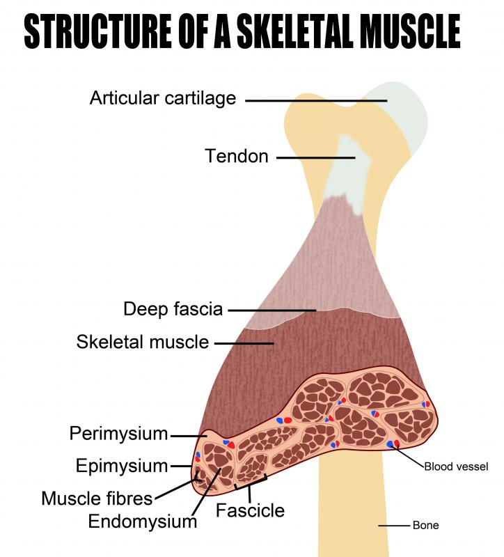
At the macroscopic level, the skeletal muscles are composed of a variety of layers. The outermost is called the epimysium, and it protects skeletal muscles from damaging friction that could occur as they move against other muscles and bones. The epimysium is a particularly important component in the anatomy of the skeletal muscles because it, along with other connective tissues, forms the muscle tendon. The muscle tendon is the strong, fibrous “rope” that keeps the muscles from slipping off their attachment points on bones.
AS FEATURED ON:
AS FEATURED ON:

-
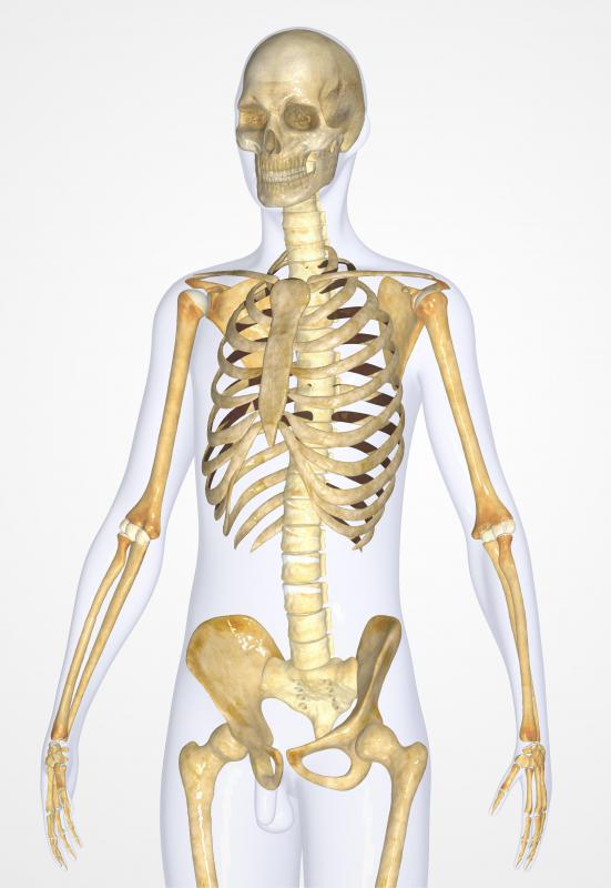
![The human muscular system.]() By: adimasThe human muscular system.
By: adimasThe human muscular system. -
![Skeletal muscle is one of three types of muscles in the body.]() By: designuaSkeletal muscle is one of three types of muscles in the body.
By: designuaSkeletal muscle is one of three types of muscles in the body. -
![At the macroscopic level, the skeletal muscles are composed of a variety of layers.]() By: Balint RaduAt the macroscopic level, the skeletal muscles are composed of a variety of layers.
By: Balint RaduAt the macroscopic level, the skeletal muscles are composed of a variety of layers. -
![The science of microscopic anatomy was discovered by Italian biologist Marcello Malpighi.]()
The science of microscopic anatomy was discovered by Italian biologist Marcello Malpighi. -
![The skeletal muscles have many layers and membranes that protect their various components.]() By: 7activestudioThe skeletal muscles have many layers and membranes that protect their various components.
By: 7activestudioThe skeletal muscles have many layers and membranes that protect their various components.


Discuss this Article
Post your comments