At TheHealthBoard, we're committed to delivering accurate, trustworthy information. Our expert-authored content is rigorously fact-checked and sourced from credible authorities. Discover how we uphold the highest standards in providing you with reliable knowledge.
What Is the Medial Condyle?
The medial condyle is a protrusion of bone that is a feature of both the femur bone in the thigh and the tibia bone in the lower leg. It is referred to as medial because in both cases it is found along the inside of the leg, toward the body’s midline. Palpable as a rounded bump on the inside of the knee in the case of the medial femoral condyle and just below the inside of the knee in the case of the medial tibial condyle, the medial condyle is in both cases a site of attachment for several major muscles and ligaments. These include the medial collateral ligament (MCL), the semimembranosus muscle of the hamstring group on the posterior thigh, and the gastrocnemius muscle of the calf.
While both bones feature a medial and lateral condyle, with the lateral condyle on the other side of the knee, the medial condyle is the larger prominence because more weight is transferred across the inside aspect of the knee joint. The paired femoral condyles are situated to either side of the patella or kneecap. They are the pair of rounded eminences that form the distal or bottom end of the femur, the largest of the human bones.

Several ligaments extrinsic to the knee joint attach to the medial and lateral condyles, connecting the femur to the tibia and fibula, respectively — most notably the paired collateral ligaments. On the side of the medial condyle is the MCL, which runs vertically to the inside of the kneecap, and on the side of the lateral condyle is the lateral collateral ligament (LCL), which runs vertically to the outside of the kneecap. Also attaching to the femoral condyles are the two heads of the gastrocnemius, the large muscle visible in the calf, with the medial head originating on the medial condyle and the lateral head arising from the lateral condyle.

Immediately below the medial femoral condyle and separated only by the medial meniscus, the disk that cushions these bones against one another at the knee, is the medial tibial condyle. Located at the tibia’s proximal or top end, it mirrors the medial femoral condyle in surface area and is a site of weight absorption on the inner aspect of the knee joint. This is where the lower end of the MCL attaches, linking the two bones together along their medial surfaces.

Also attaching to the medial tibial condyle is the tendon of the semimembranosus muscle, the innermost muscle of the hamstring group on the posterior inner thigh. This muscle contributes to flexion or curling of the knee by pulling upward on the tibia bone and thereby causing the joint to hinge. On the other side of the knee, the most lateral of the hamstring muscles — the biceps femoris — crosses the joint and attaches to the lateral tibial condyle much as the lateral head of the gastrocnemius connects to the lateral femoral condyle above.
AS FEATURED ON:
AS FEATURED ON:

-
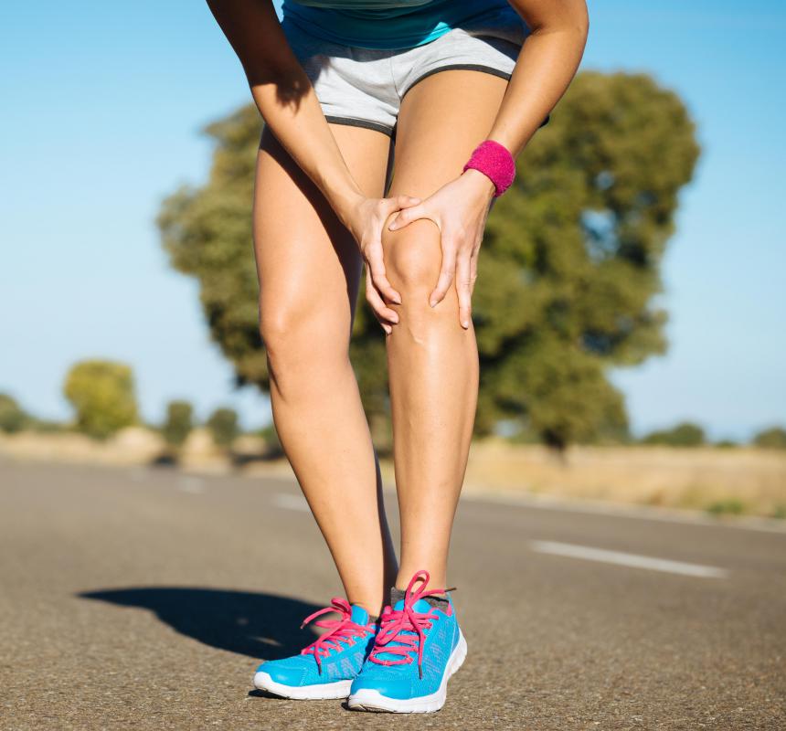
![The medial collateral ligament (MCL) runs vertically to the inside of the kneecap.]() By: OOZThe medial collateral ligament (MCL) runs vertically to the inside of the kneecap.
By: OOZThe medial collateral ligament (MCL) runs vertically to the inside of the kneecap. -
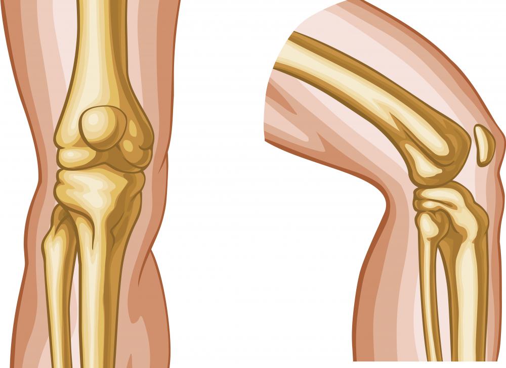
![A diagram of the knee, showing the medial condyles, where the medial collateral ligament attaches to the femur and tibia.]() By: AlilaA diagram of the knee, showing the medial condyles, where the medial collateral ligament attaches to the femur and tibia.
By: AlilaA diagram of the knee, showing the medial condyles, where the medial collateral ligament attaches to the femur and tibia. -
![The medial condyle is a feature of the femur bone in the thigh.]() By: maya2008The medial condyle is a feature of the femur bone in the thigh.
By: maya2008The medial condyle is a feature of the femur bone in the thigh. -
![Most people refer to the patella as the kneecap.]() By: DirimaMost people refer to the patella as the kneecap.
By: DirimaMost people refer to the patella as the kneecap.


Discuss this Article
Post your comments